
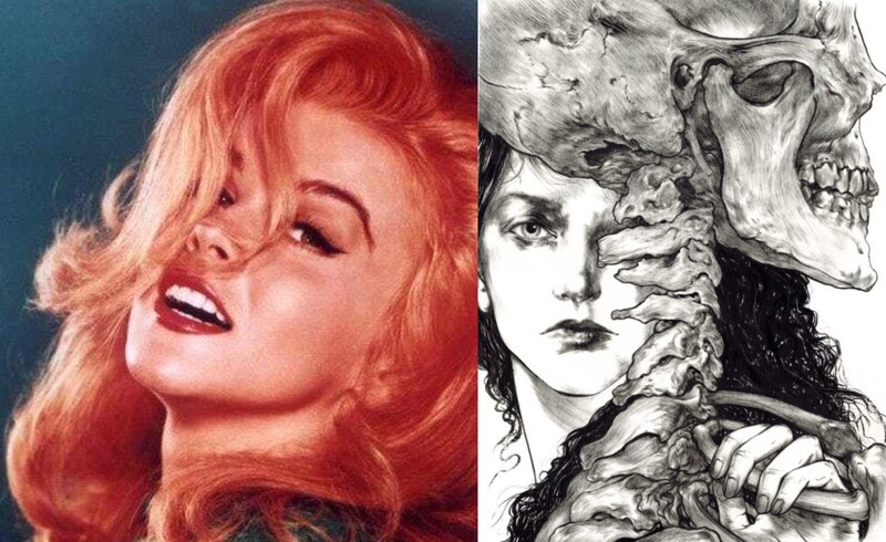
The internet is full of cries for help: headaches, unsteadiness and nervousness appeared after braces; ‘they ruined my face, neck and spine’. This is only a tiny fraction of the problems that arise, but alas, these complaints drown in tonnes of bright advertising for Hollywood smiles with braces. Nor do they find understanding from the orthodontists who fitted those braces: they try to get rid of such ‘problem’ patients as quickly as possible, telling them it is psychological or, worse still, simply laughing at them.
Unfortunately, this is not funny, because the harm braces cause a patient is sometimes comparable to a traumatic brain injury, as we will see below. I regret not knowing about these consequences earlier and having to fill the gaps in my orthodontist’s knowledge myself as a patient. This article brings together information from recognised world-class TMJ specialists, craniodontists, orthopaedists, posturologists and osteopaths: for example, the legendary early-twentieth-century American orthopaedist Robert Lovett, famous osteopaths Horace Magoun and Viola Frymann, the brilliant Gerald Smith, Darick Nordstrom, Guiseppe Stefanelli, Leopold Busquet, John and Mike Mew, and others. Also included is Dr A. O. Savinov, the founder of orthocraniodontics in our country, who provided most of the references and explanations on his former orthocranio.ru forum and forum.stom.ru.
Based on these doctors’ research, let us examine how braces can affect the skull, neck region, spine and overall health. If you recognise any of the things listed here after having braces, do not despair: as I have been able to establish, there is a way out of this situation too, which we will discuss below. We will also examine why many patients do not have these health consequences and in what cases braces can still be used. Now let us turn to the main point: the crippling consequences traditional braces treatment can have for a patient.
1. The Face

In the article on how braces damage the face, we examined in detail how braces draw out and lower the maxilla, the ‘beauty bone’ responsible for forming the midface. The photograph above shows another clear example of this negative effect on beauty: braces fitted to the girl at twelve meant that by fourteen her face had lengthened and flattened, losing its horizontally developed three-dimensional structure. Drawing the maxilla downward also meant that her eyeballs lost support from below, producing a ‘bulging eyes’ effect. But extracting teeth for braces has still more catastrophic consequences for facial beauty.

In the first photograph above, we see the face of a woman who had two upper-arch teeth removed during previous orthodontic treatment for braces. Her face became simply ‘crumpled’ and folded inward, because skin and muscles always cover the skeletal structure like fabric over a frame! Fortunately, the patient found a competent craniodontist working with ALF. Her upper jaw was expanded, space was created for the extracted teeth (the intermediate photograph), and implants were placed. In the final photograph we see an attractive, three-dimensionally developed face. The patient also looks about ten years younger! Compare the final photograph again with the first, where her teeth had been removed and the spaces closed with braces. Unfortunately, losing facial beauty is not the worst thing that can await a patient after braces, especially when teeth are extracted for them…
2. The Skull
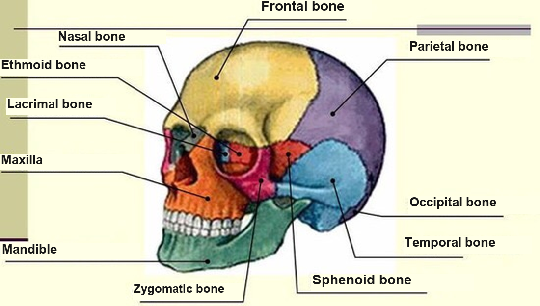
The Straight Wire Appliance (SWA), or simply braces, is merely a cosmetic procedure aimed at improving the appearance of the dental arches, mainly at the front. Unfortunately, orthodontists often forget that the body’s stomatognathic system includes, besides the teeth: 1) the facial and cranial bones; 2) the joints of the head; and 3) the neuromuscular complex of the head and neck.

There is also the craniosacral system, whose most important component is the so-called craniosacral mechanism (CSM), or primary respiratory mechanism. It is called a mechanism because it manifests through the complex articulations of the cranial bones — through mechanics. The CSM is considered the basic mechanism of life; a conflict at one level leads to various dysfunctions and subsequently persistent chronic diseases that even specialist doctors often cannot diagnose. Braces restrain the physiological mobility of the cranial bones, thereby blocking the craniosacral rhythm.
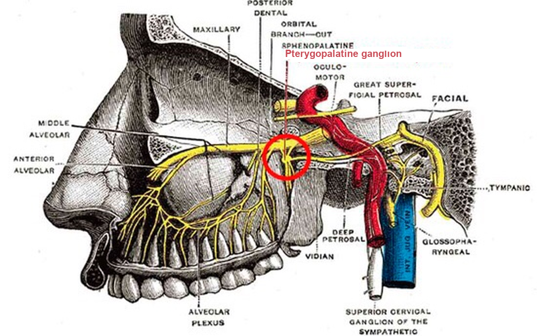
Symptoms associated with disturbances of the craniosacral mechanism can be very varied. For example: 1) vasomotor rhinitis caused by blockage of the palatine, ethmoid and sphenoid bones and upper jaw (maxilla), disrupting drainage of the ethmoid cells and opening of the nasal conchae; 2) compression and irritation of the pterygopalatine ganglion, formed from a branch of the trigeminal nerve, causes persistent facial pain and autonomic nervous system disorders; 3) twisting of the sphenoid bone deforms and narrows the superior orbital fissure, affecting the oculomotor, trochlear, trigeminal and abducens nerves and causing ophthalmological problems and even strabismus;

4) distorted rotation of the temporal bones affects the internal auditory opening and vestibulocochlear nerve, leading to hearing problems and unstable balance; 5) cranial distortions affecting the diaphragm of the sella turcica inside the skull cause hormonal imbalance and affect women’s menstrual cycles, PMS and reduced libido in women and men through their effect on the pituitary gland; 6) compression of the occipital bone compresses the cerebellum, impairing coordination and causing unsteadiness, and also indirectly affects the pituitary gland; 7) any displacement near the occipital opening compresses the nerve plexuses of major vessels and arteries entering the cranial cavity through the jugular foramen, irritating the brainstem and vagus nerve. Hence headaches and migraines, breathing problems, gastrointestinal problems, constipation, diarrhoea and restless behaviour.

All of this was presented not to tire you, but to show the scale of destruction that even the smallest intervention in the body’s stomatognathic system can cause. The most unpredictable consequences occur when ‘extra’ teeth are extracted and elastics are used, because this sets all twenty-two pairs of cranial bones in motion! Remember that most patients already have disturbances of the craniosacral mechanism when they visit an orthodontist. I do not want to paint too dark a picture, but now you understand why some cases after braces are comparable to a traumatic brain injury, after which, alongside the main treatment, a person needs lengthy rehabilitation — not a psychologist, as many orthodontists try to send you to in this condition! These are very serious matters.
3. The Temporomandibular Joints

One of the most frequent injuries caused by braces! From the above, we have seen how braces can affect the entire cranial structure. The temporomandibular joints lie at the intersection of this three-dimensional system, and displacement in even one plane inevitably displaces the joints and discs too. Roughly speaking, if we compare the skull to a little house tilted to one side, it is not hard to imagine what happens to its door hinges (the temporomandibular joints). But this is only one possible displacement.

The ‘house’ can also tilt forward or backward and ‘turn’ slightly around its axis. I think there is no need to describe what happens to the door hinges (the temporomandibular joints). The symptoms experienced by the ‘lucky ones’ whose skulls underwent this can be found in all their ‘colourful’ detail in the article on TMJ, the ‘chameleon disease’. Here I again want to emphasise that patients often come to an orthodontist with existing cranial distortions, and thoughtless intervention with braces only greatly worsens the process. We will consider the treatment algorithm a little later. For now, let us examine how damage at the cranio-mandibular level triggers deformation of the skeletal structures below.
4. The Atlas and Cervical Vertebrae

The picture shows the simplest diagram of this effect. As we see, with a horizontally developed, forward-positioned maxilla (upper jaw), the skull is in a physiological upright position, supported at the front by the lower jaw (more detail on this biomechanical process is in this chapter of Moreno Conte’s book ‘How I Straightened My Spine’). In the article on how braces damage the face, we examined how braces pull the maxilla backward and downward. This also blocks movement of the lower jaw, forcing it to move along a newly prescribed path. Consequently, the head inevitably drops forward, causing changes in all cervical vertebrae by a ‘domino effect’, beginning with the first, the atlas. Yet this is far from everything, since we have considered the skull’s dropping only in the lateral plane. There are also the frontal and transverse planes! Remember that the human skull is a three-dimensional system:

If braces damage this system against a background of existing cranial distortions, the skull may tilt not only forward but also to one side and twist around its vertical axis. You can imagine what happens to the atlas, which always adapts to the occipital bone’s position. Many also know firsthand the consequences of displacement of the first cervical vertebra (alas, I do too). The atlas position greatly affects a person’s nervous and energetic systems and ability to feel happiness. If you are considering braces, think about this too. You now also understand how dropping and twisting of the skull lead to cervical scoliosis and lordosis. With congenital cranial deformities, this process takes years, giving the body time to develop adaptive mechanisms. With braces, especially tooth extractions for them, it develops fairly quickly and painfully, because such an abrupt intervention shocks the body.
5. Neck Muscles and the Vertebral Artery

As the skull drops forward and cervical lordosis develops, the neck muscles at the back shorten while those at the front lengthen. Cervical scoliosis worsens this situation: muscular chains running from the lower jaw through the hyoid bone to the clavicles, and from the occipital bone to the clavicles, develop tension of different intensity on different sides. Now you can imagine what happens to the vertebral arteries feeding the brain: they run partly through the bony canals of displaced cervical vertebrae and partly under compression beneath the scalene muscles.

For the full picture, let us also remember the cervical plexus, formed by the anterior branches of the four upper cervical spinal nerves. It lies on the surface of the deep neck muscles (levator scapulae, medial scalene and splenius) at the level of the four upper cervical vertebrae. They innervate all nearby neck and head muscles and give cutaneous branches of the great auricular and lesser occipital nerves. The cervical plexus’s phrenic nerve also descends along the anterior surface of the scalene muscle and enters the thoracic cavity through the upper thoracic aperture, passing close to the pericardium and ending within the diaphragm. Can you imagine what happens when overstrained neck muscles compress it? This too is a direct consequence of braces! But that is still not everything.
6. The Spine and Pelvis

As early as the beginning of the twentieth century, orthopaedist Robert Lovett described S-shaped and #-shaped relationships within the human skeleton. In these, the occipital bone correlates with the thoracic spine (T1–5), lumbar region and sacrum, while the temporal bones correlate with the cervical spine (C1–6), shoulder blades, ribs, thoracic spine (T6–12) and pelvic bones. Thus, any change in the occipital bone’s spatial position changes the upper thoracic and lumbar spine and sacrum (a reverse relationship can also be observed). Asymmetrical rotation of the temporal bones always occurs simultaneously with asymmetrical rotation of the pelvic (iliac) bones — synchronously but as a mirror image — and with changes in the cervical and lower thoracic spine.

Such skeletal changes after braces do not occur all at once, because the relationship here is linear rather than direct. But after just a few short months, patients may notice the first signs of these disturbances, which develop over years, gradually changing all the skeletal and muscular structures below, down to the feet…
Not ‘Who Is to Blame?’ but ‘What Can Be Done?’
Einstein said a problem cannot be solved at the level where it arose, only by moving one level higher. A different approach is offered as an alternative to braces: before fitting a Straight Wire Appliance — simply, braces — existing cranial deformities must be corrected. Correcting them begins with stabilising the temporomandibular joints and correcting the maxilla’s position using the intraoral ALF appliance, integrated with osteopathy. This method, called orthocraniodontics in our country, aims to stimulate self-correction of dentofacial deformities; braces are introduced only at the final phase, to affect, for example, the axial position of the front teeth.

After cranial deformities are corrected, braces do not disturb the craniosacral mechanism, because this is guaranteed by the philosophy and method of the functional ALF appliance, which can work simultaneously with braces. Here lies the answer to why many patients have none of the negative health consequences listed above after braces (except perhaps complaints about an altered face), while for others braces ‘treatment’ is quite crippling. It depends precisely on the initial state of the cranial system and existing deformities with which the patient comes to the orthodontist. The other question is: how many patients come without cranial and skeletal disturbances?

The dentofacial system’s own adaptive capacity is also quite considerable, allowing a thoughtless dentist to violate nature mercilessly. So although traditionally practising orthodontists give, to put it mildly, insufficient attention to cranial distortions and joints, some patients’ braces ‘treatment’ still goes more or less smoothly, especially when young — and for some, not quite… I became one of those patients. Since creating this blog, many people have written to me with far worse situations: after braces, especially with extractions, they lost not only their health but their work, friends, and some even their families and faith in themselves. Orthodontists must therefore rethink what they do, because sometimes not only a patient’s health but their future fate depends on the quality of their work. Let us leave questions of karma, cause and effect for now — we will discuss them another time.
The most upsetting thing is that timely, completely free myofunctional exercises could have prevented the need for an orthodontist altogether. They restore dental-arch width and facial beauty, also completely curing TMJ problems (according to British orthotropics doctor Mike Mew and Norwegian craniodontist Geir Olsen). But this is not about severe cases following braces, particularly tooth extractions and elastics. ALF treatment is an alternative in such cases, but unfortunately is no picnic either: during this process absolutely every previous symptom returns in reverse order, and great strength of spirit is needed to endure the treatment. Despite all orthocraniodontics’ merits, another major obstacle is the very small number of truly high-level practitioners in our country. Finding oneself in this profession requires more than simply taking courses: one must keep studying for many years, not only ALF orthodontics but osteopathy, posturology, orthopaedics and other related specialties. Here the doctor needs strength of spirit too. Nevertheless, I sincerely believe the future of orthodontics as a science lies in this field and its close relationship with cranial osteopathy.
Update, 4 December 2021: After running this blog for four years, I regret to report that I receive differing reviews of practitioners using intraoral appliances: some patients recover with the same doctor, while others become much worse than before treatment. I follow the situation objectively, comparing these reviews with those received over the last two years about the use of finger techniques. Most of these people had suffered harm after braces. Essentially, the method is self-osteopathy: its techniques not only decompress facial and cranial bones but also considerably align the jaws relative to each other, which affects the bite too .
Two women shared their experiences. One had a traumatic brain and facial injury in a road accident. She completely restored her face and, more importantly for our topic, her neurological symptoms, migraines and depression disappeared (unfortunately, I cannot publish her photograph here). The second woman, whose photograph you see below, had cranial surgery four years earlier and half her face paralysed. The second photograph shows her after three months of finger techniques, with noticeable progress in facial-muscle movement!

And this correction was undertaken by the women as an experiment and a last hope: the method’s author honestly told both that she had never encountered such cases and did not know whether it would help. Perhaps this information may be useful to some of you. I too cannot take responsibility for recommending the method in severe conditions, so responsibility for your choice rests with you. I am simply honestly adding to the information I have collected.
Orthocraniodontics: Philosophy and the Basic Treatment Algorithm — here.
__________________
5 comments
Ask a careful question or add useful context. New comments are reviewed before publication.
“Therefore, any change in the spatial position of the occipital bone leads to changes in the position of the upper thoracic and lumbar spine and sacrum”
The question is how is it all connected to the mewing and the development of the maxilla? And does the mandible growth also effect it position of the occipital or just the maxilla?
What the more the maxilla grows forward and upward or the mandible hoe exactly it effects the occpital bone?
There is not enough information about this i want to read more …
Hope you will replay