
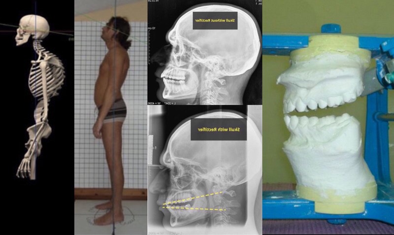
This article shows that posture is directly related to the correlation between the skull and jaw, and that if your teeth are not symmetrically positioned, you are more likely to experience postural problems such as scoliosis, lordosis, kyphosis, body asymmetry, and musculoskeletal twists.
In this chapter of the book “How I Straightened My Spine”, the scientific theory behind the functioning of postural biomechanism has already been examined. In short, we saw that this mechanism is based on the relationship between the skull and the jaw, and it is this relationship that determines the position of the human body.
We also realized that the position of the skull on the jaw depends on the symmetrical height and position of the teeth on both sides. Finally, we have seen that if the occlusal plane is staggered or asymmetrical, an incorrect correlation between the skull and mandible can have a negative effect on the position of the whole body.
In simple terms, the skull literally rests on the first cervical vertebra atlas, and the dental arches are the “scaffolding” that supports the skull. We also saw from the book above that this support is temporary and appears during the closing of the teeth. During occlusion, through the swallowing mechanism, the structure of the neuromuscular interaction between the skull and the jaw is transferred to the underlying structures.
In fact, the weight of the skull is distributed over the teeth – for example, the molars are larger because they discharge most of the weight of the skull. Conversely, the anterior incisors are thinner because they do not need to support the skull and only serve to cut food.
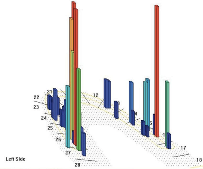

In fact, the tooth that is most stressed by the weight of the skull is the first large molar (the molar at the back of the jaw). It is not a coincidence that on the upper and lower dental arches molars are present in descending order – the first is more than the second, the second is more than the third, etc. In fact, this is how the weight of the skull is distributed.
Therefore, if the large teeth do not have sufficient height, the skull can no longer be supported on them and sinks, exerting compression on the spine, which, in turn, becomes compressed in a small space. It is this compression that leads to the phenomena of kyphosis, lordosis and scoliosis, and causes compression of nerves, muscles, internal organs, arteries, cartilage, etc. It is these compressions that cause numerous symptoms and even lead to chronic diseases.
The interaction between teeth, their closing, the effect on the spine, and various disease symptoms have been hypothesized and explored by several branches of science in recent decades. But no one could clearly understand this mechanism in order to stop the process of destruction of the body in the most decisive way.
In this article, we intend to finally demonstrate the empirical evidence that the position of the body depends on the teeth. But before embarking on this demonstration, below are three scientific articles that will help us understand the current state of scientific research on the relationship of occlusion, posture and emerging disease symptoms:
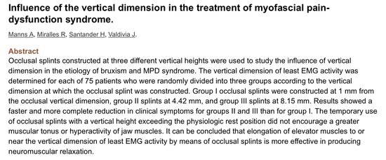
1) the first article is dated November 1983 and shows the results of a study conducted by the Medical University of Santiago (Chile). For this study, 75 patients were selected (9 men and 66 women, ages 13 to 53);
2) the second article is about the relationship between teeth and body position of some rodents;
3) the third article analyzes the effectiveness of the occlusal splint to improve postural structure and athletic performance.
Scientific article No. 1
http://www.ncbi.nlm.nih.gov/pubmed/6580439
The patients were divided into three groups:
Group 1: 25 patients made an increase in the vertical height of teeth by 1 mm (3 men and 22 women aged 13 to 52 years) Group 2: 25 patients – an increase of 4.42 mm (4 men and 21 women aged 15 to 52 years old) Group 3: 25 patients – an increase of 8.15 mm (2 men and 23 women aged 15 to 53 years)
Each of the participants in the experiment was required to insert a flat splint into the mouth for 3 hours a day for 3 weeks. During this period, the manifestation of twenty-one types of psychosomatics was studied. Each trait was scored on a 0 to 3 scale (0: not present, 1: significant improvement, 2: slight improvement, 3: still present) Symptoms were monitored for 24 h, 72 h, 1 week, 2 weeks and 3 weeks after the change bite.
The aim of this study was to test whether tooth height can influence the solution of problems such as bruxism and identification disorders (depersonalization, derealization, etc.) and this is what was found:
The first interesting data is that immediately after 72 hours, groups 2 and 3, where a significant increase in bite height was made, showed a 50% reduction in symptoms. In contrast, group 1, in which a 1 mm increase in tooth height was performed, showed the same improvement after a week. Overall, patients in groups 2 and 3 took much less time to reduce or completely resolve symptoms than patients in group 1.
It was also found that increasing the bite height between 4 and 8 mm not only increased muscle work, but also decreased muscle tension. The increase in bite in this case, on the contrary, relaxed the muscles.
Groups 2 and 3 had more than 70% improvement in symptoms such as neck pain, headache, TMJ pain, pain in the face and skull bones, morning and evening muscle fatigue, tinnitus, dizziness and some others. Also, the condition of the previously deflected lower jaw in many patients has significantly improved, clicks in the temporomandibular joints, limited mouth opening and nocturnal bruxism have decreased. Thus, these studies confirmed that the height of the bite has a direct effect on the main structures of the body, for example, the neck, while the symptoms associated with changes in this area were reduced by 70%.
Scientific article No. 2
http://www.ncbi.nlm.nih.gov/pubmed/25046977
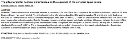
This study, conducted in mice, demonstrated that removing teeth from these rodents or placing overly high fillings in their teeth altered the curvature of their spine.
![Source image captions: The effect of dental occlusal disturbances on the curvature of the vertebral spine in rats.; Ramirez-Yanez GO, Mehta L, Mehta NR.; Abstract; Objective: To determine whether a unilateral increase or decrease in the bite affects the curvatures of the vertebral spine in rats. Methods: 25 male Sprague Dawley rats were included. Five animals received no alteration on their bite. Bite was increased on 10 animals and molar teeth were extracted on 10 other animals. Frontal and lateral radiographs were taken on days 0, 7, 14 and 21. Distances from landmarks to a true vertical line were measured on both radiographs. Results: Repeated measures analysis showed statistically significant differences between the amount of the curvature at the cervical and thoracic spines on frontal and lateral radiographs over time (P<0.05 and P<0.0001). One-way ANOVA computed significant differences (P<0.05) at D14 at the cervical and thoracic spines in both, increased bite and decreased vertical dimension. Conclusion: Alterations in the dental occlusion affects the normal curvatures of the vertebral spine in rats.; KEYWORDS: Body posture; Dental occlusion; Mandibular function; Physiological curvature; Vertebral spine.; PMID: 25046977 [PubMed - as supplied by publisher]](https://media.jawandbody.com/assets/article-images/english/alf092-source-captions-rat-spine-abstract-en-v1.svg)
Scientific article No. 3
http://www.ncbi.nlm.nih.gov/pmc/articles/PMC3476490
This study confirms that wearing a special dental splint can improve the performance of athletes by increasing their quadriceps strength. In addition, the use of a dental splint eliminated pain in the lumbar region. This study was carried out by a group of gnathologists after basketball team doctors were unable to solve the problems of one of the athletes with conventional physical therapy. A special splint was made for the athlete, and his symptoms immediately decreased and his muscle strength increased.
But, despite all these studies, none of the scientists has established unequivocally that posture depends on the bite, or rather, on the cranio-jaw relationship. Moreover, no effective practical solution has been proposed for postural problems in their connection with the bite. We can say that something happened with the advent of the Italian Starecta method, which made it possible to radically influence the regulation mechanism of the biological body mechanism. These results were achieved through the development of a new methodology that can be applied and regulated individually. Starecta is fundamentally different from previously used methods, which turned out to be incomplete.
Typically, these methods used an expensive piece of dental resin that proved useless in the long run. But now, after years of searching in the dark, there is finally an answer. We have data to support previous scientific research. The correlation between bite and posture has been demonstrated by the experience of a person using the Starecta method.
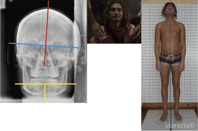

Former professional footballer of the Italian club Civitanova Simone Cincini had an undiagnosed cranio-jaw imbalance problem, like 98% of people. Before solving this problem with Starecta, this patient had tried every possible treatment.
Let’s take a look at Simone’s problem as identified by the Starecta team. Simone had:
– lateral deviation of the skull to the left, which created a significant asymmetry of the musculoskeletal system in the frontal plane;
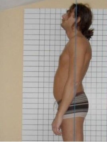
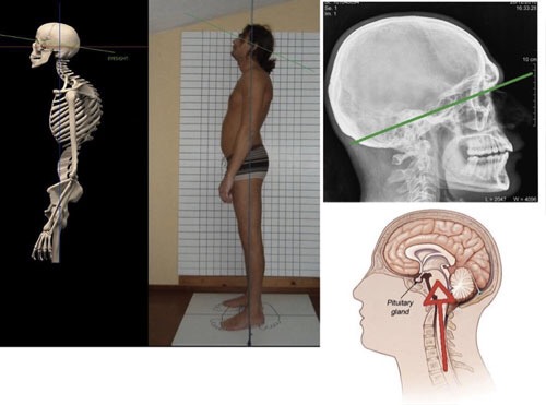
– a huge lack of dental height in the lateral (sagittal plane) on both dental arches in the area of the large and small molars, which caused an increase in thoracic kyphosis, and cervical and lumbar lordosis, the eyes looked up.
By looking at an X-ray of Simone’s skull in the frontal plane, we can see that the lack of sufficient dental height on the left dental arch caused a deformity of the skull at this point. Simone, like many of us, was probably born with this asymmetry, which only got worse over time, making his life nearly impossible. Lateral deviation of the skull to the left caused facial and then body asymmetry.
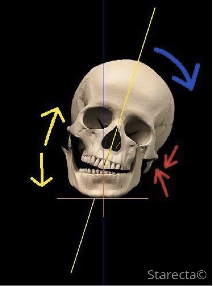
With such a lateral deviation of the skull, the chewing muscles, and then the temporal muscles, become shorter, and on the other hand, they lengthen. Also, we all know that when a muscle shortens, it becomes stronger, and in a stretched state, it becomes weaker. Thus, in the case of lateral deviation to the left, the muscles on this side are much more developed and pull in their direction all the soft tissues, bones and cartilage. Knowing this, it is easy to understand why Simone’s nose deviated to the left for many years, his left eye dropped, and his left cheekbone developed more than the right one. The lateral deviation of the skull to the right then spread to the rest of the body, causing musculoskeletal asymmetry, twisting of the spine, pelvis and legs.
Simone’s biggest problem is in the lateral (sagittal) plane – since he had a significant lack of dental height in the area of the large and small molars, his skull literally “sagged” in this area – he fell back and turned up. This position of the skull caused compression of the spine, which was forced to be in a reduced compressed space. That is why Simone showed an increase in the curvature of the normal physiological curves of the spine, which began to appear in the form of kyphosis and lordosis, as we see in the photograph.
If this is not enough, then it is important to note that the spinal cord runs inside the spine. “Falling” of the skull backward created compression of the spinal cord, which was already causing serious symptoms. Looking at the picture below, try to imagine what is happening to the skull, and therefore to the spine and spinal cord, if you remove the red triangle located between the dental arches.
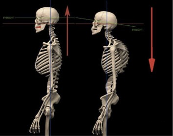

The red triangle is the ideal height of the teeth in the area of the large and small molars, and thanks to it, the skull is kept in an optimal position. Therefore, if we remove this red triangle (creating the ideal bite height in the area of the large and small molars), the skull will change its slope, turning upward until it finds a new fulcrum in the area of the large and small molars. As we understood earlier, this position of the skull exerts compression on the spine, forcing it to change its physiological curves, as can be seen in the photo below.

Compression of the spinal cord also creates pressure on some areas of the brain. During compression, the spinal cord resists the sinking effect of the skull and pushes the lower brain upward. The problem lies in the fact that between the cranial and spinal cord is the pituitary gland, which begins to undergo increased compression.
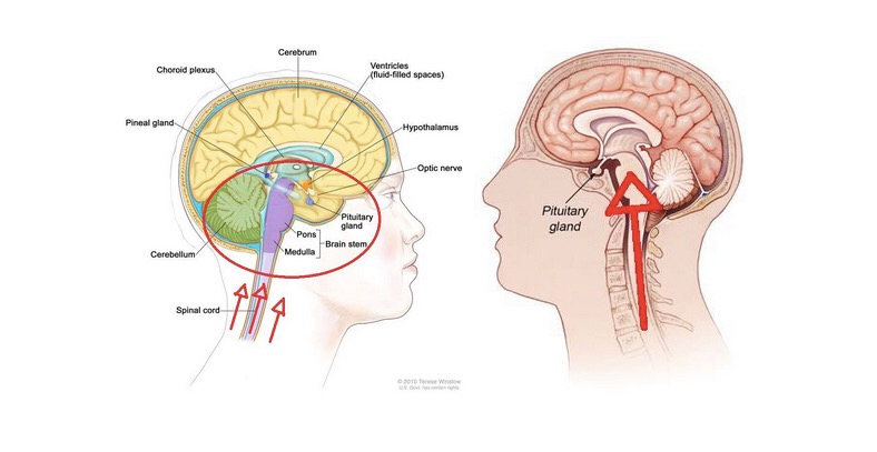
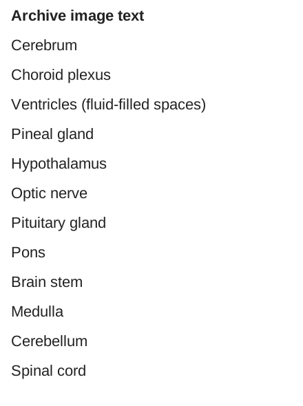
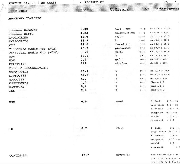
This pituitary gland, located in a pocket of bone called the Turkish saddle, is the central organ of the endocrine system and secretes hormones that affect growth, metabolism, and female and male reproductive functions. The hormone gonadotropin produced by the pituitary gland is responsible for the regulation of the work of the gonads. If its values in the analyzes are equal to zero, this means that the pituitary gland is completely blocked. This is exactly what happened to Simone. His spinal cord and pituitary gland were compressed by a sunken skull. This blockage of the pituitary gland was caused by a cranio-mandibular imbalance – little known and therefore undiagnosed.
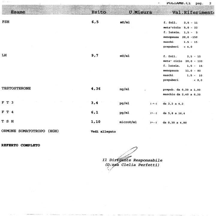
Below are medical tests that support this fact. As you can see in the pre-Starecta test, the gonadotropin hormone (LH and FSH) values are almost zero (0.2 and 0.0 respectively). After applying the Starecta method and returning the skull to the optimal position, these values returned to normal (9.7 and 6.5, respectively).

Below we will see how biomechanical and biochemical mechanisms are intertwined in the body. Why do hormone levels return to normal after using Starecta? This method uses a tool that can stop and reverse the sinking and sinking of the skull, returning it to its ideal position. This allows us to solve the problems of compression of the vertebral structures, which we talked about earlier.
In practice, the intraoral device of the Starecta method is inserted between the dental arches and, according to a certain methodology, directly affects the biomechanism that regulates the position of the skull and provides decompression of the compressed areas of the brain.
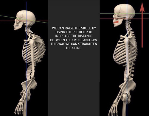
As you can see from the above medical tests performed before and after the application of the Starecta method using the Rectifier splint, the position of the skull was stabilized, the spine was decompression and the release of the pituitary gland, which resumed its functions.
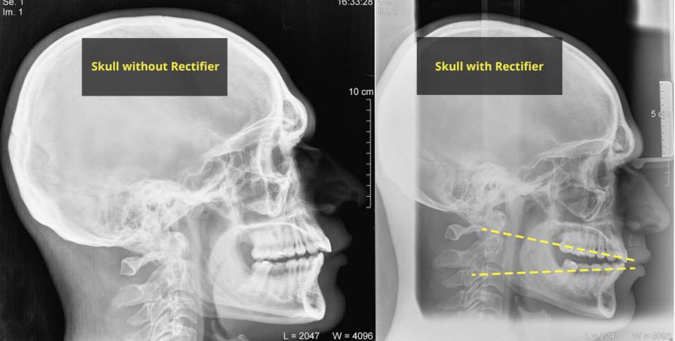

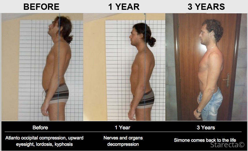
We can distinguish three elements that demonstrate the uniqueness and scientific basis of the Starecta method:
1) Simone Cincini’s photographs clearly demonstrate how his posture was changed, which seemed impossible before the method was applied;
2) X-rays of the skull and cervical vertebrae show that the skull actually rose due to the space that was created by the Rectifier. This space is the missing ideal bite height necessary for the skull to maintain its correct physiological state. This space has been restored thanks to the Rectifier;
3) medical tests demonstrate that prior to the application of the method, the pituitary gland in this patient was completely blocked due to cranio-jaw imbalance, which was not previously diagnosed by doctors. It was only after the application of Starecta that the pituitary gland resumed its normal functioning.
The Starecta method is quite young and this article is one of the first scientific evidences to demonstrate its effectiveness. But over the coming years, the scientific evidence for the method will increase at a growing progression.
________________________


6 comments
Ask a careful question or add useful context. New comments are reviewed before publication.
What should you do about occlusion after Starecta? (available later)
Everything is written there in detail.
Starecta Rectifier
You should also know that, according to the method’s technology, the effect is on the posture below, but when it comes to correcting skull deformities, the splint is somewhat limited, and it is preferable to add mewing to its effect. This is described in great detail, with the story and experience of one of our guys, here:
Starecta and mewing: how to reconcile conflicting forces (available later)
Or you can go straight to a craniodontist who works with all of these issues at once. There are such specialists in my area, and we have a list of them here:
Craniodontics: the best specialists
We are trying to collect reviews of doctors in the discussion section here:
You can find related materials in this archive.
Quite a few people are already being treated by these specialists, and you can talk to them in our reader discussion area:
You can find related materials in this archive.
I really hope all this information will help you get your bearings. And I wish you recovery!
If you have any more questions, write.