
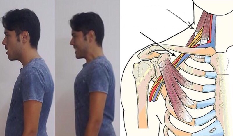
This article is not scientific. In it, I merely want to share my observations of the ‘behaviour’ of the vertebral arteries when the position of the head and neck changes; the conclusions are yours to draw. Whether compression of the vertebral arteries by the deep neck muscles and in the subclavian region disappears in this case, whether their diameter is restored after that compression is eliminated, and how straightening posture affects this entire process — I think many people would find it interesting to understand this.
Experimental results of Starecta and orthocraniodontics
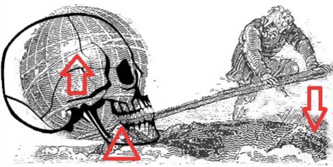
On this blog, I have already described the biomechanical principle of the spine-straightening method Starecta and the mind-boggling possibilities of orthocraniodontics. Unfortunately, there are very few accounts online from people who have undergone treatment using the latter method; I have managed to speak to only a few such patients. I suspect this is because, having climbed out of hell and enjoying the long-awaited, hard-won feeling of health, a person wants to forget it all as quickly as possible and no longer feels like sitting on the internet; the superstitiousness of the Russian character also plays a part (I am like that myself). Foreigners, however, are more open in this respect, and the Starecta method is currently better known worldwide.
Following accounts by people using Starecta, I cannot help sharing my observations of the results of this method in people with symptoms typical of vertebral artery syndrome: dizziness, chronic fatigue, lack of concentration, needing a great deal of sleep and ‘brain fog’. It should be noted that they generally do not have their vertebral arteries examined, since doctors around the world have difficulty diagnosing vertebral artery syndrome and thoracic outlet syndrome (yes, patients in the post-Soviet region are not alone in this respect). As a rule, these patients, like ours, are prescribed massages, physiotherapy, muscle relaxants and so on. They are sent to psychologists too. But after the first days of using this experimental method, they generally write the following:


Today is my first day with the Rectifier. Usually, all day long I feel anxiety, pain in my neck and the back of my head, pressure in my head, severe fatigue, imbalance/a feeling of dizziness, numbness in my right arm, poor vision and black spots in my right eye, cramps, spasms and tingling all over my body, as if an electric current were passing through me, ringing in my right ear, slurred speech… I took my daughter to school today and, by the time I returned, I was covered in sweat and felt intense anxiety… severe pain in my head; I nearly fell twice. At 10 a.m., I put the Rectifier in my mouth and… 30 minutes later, I felt my right ear open up; it was strange, but good. An hour later, I noticed that I was walking without feeling that I was about to fall. I went to the school: no anxiety, no feeling of falling and no dizziness… I can move my head without severe pain. I have not taken painkillers today. It is now 9 p.m., and I am still awake, whereas I usually crawled into bed at 6.30…
Or:

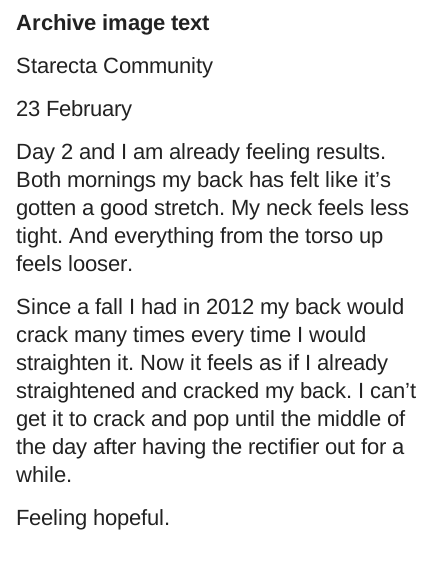
Day 2, and I already feel results. For two mornings in a row, I have felt a pleasant stretching in my back. My neck has become less tense. My torso feels freer…
A similar effect occurs when orthocraniodontic techniques are used (though not immediately). But what happens to the neck, vertebrae and vertebral arteries in the process? Why do people feel results instantly? To answer this question, let us turn to some slightly ‘tedious’ theory.
Vertebral artery syndrome, the cervical outlet and the thoracic outlet
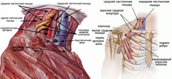
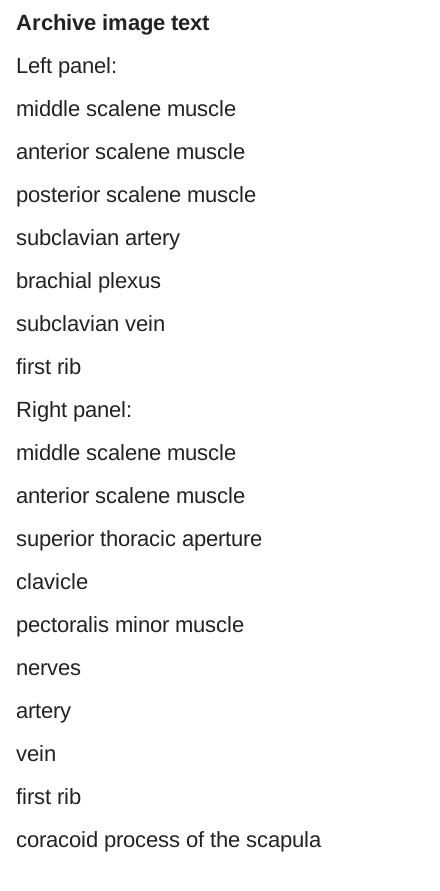
In his, without exaggeration, magnificent works ‘Vertebrobasilar Insufficiency’ and ‘Thoracic Outlet Syndrome’, which many of us know well, Lithuanian vascular surgeon Povilas Pauliukas describes compression of the vertebral artery by the deep neck muscles (in particular, the scalene muscles), as well as compression of the subclavian artery between the anterior and middle scalene muscles (cervical outlet syndrome) and compression of that same artery in the costoclavicular space (thoracic outlet syndrome). In these cases, he proposes surgery to eliminate the compression. At the same time, Povilas Pauliukas also writes that poor posture can cause arterial compression in thoracic outlet syndrome, since it reduces the space between the clavicle and the first rib. As we see in the animation below, this phenomenon does indeed occur with poor posture. Moreover, the more pronounced the cervical hyperlordosis and thoracic kyphosis, the less space remains between the clavicle and the first rib.
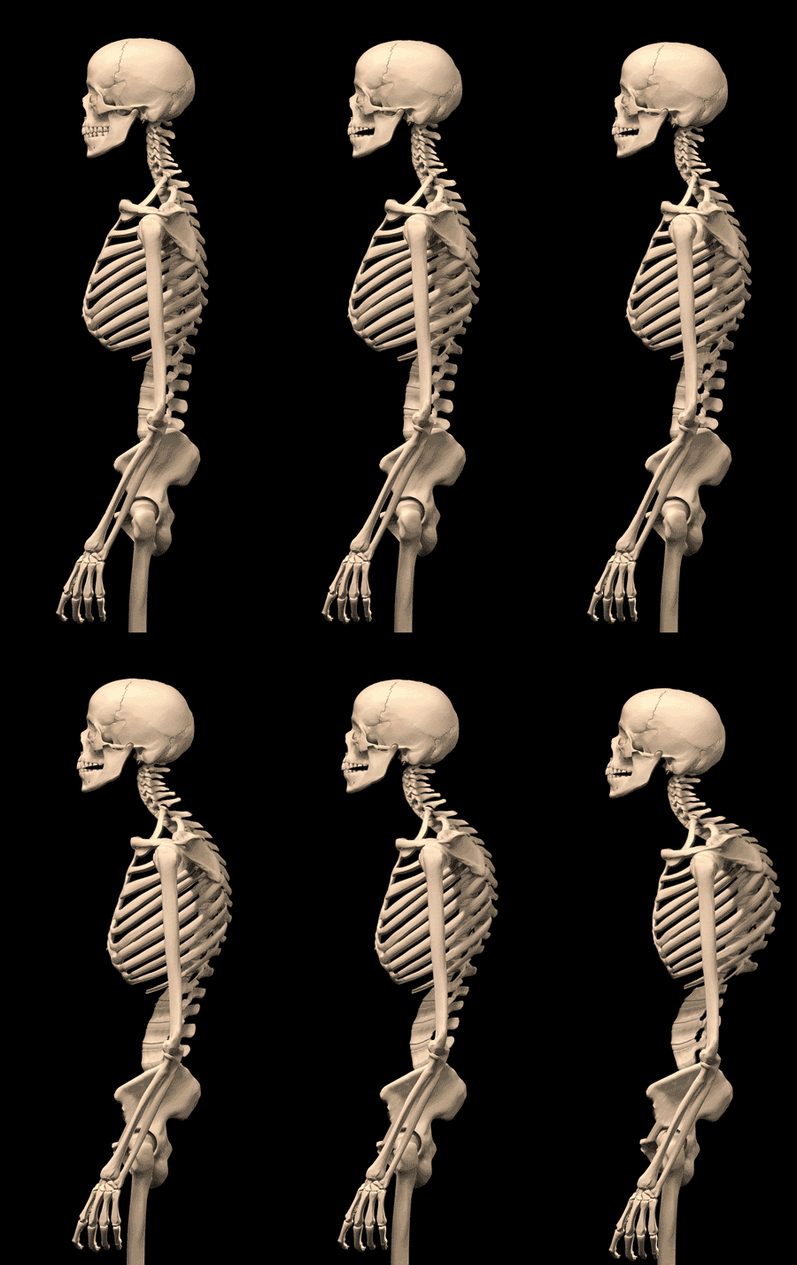
It is not difficult to guess what happens to the subclavian arteries in this situation. Perhaps this also explains compression of the vertebral arteries at their origins in the lower part of the neck. But what happens to the vertebrae and neck muscles with poor posture and loss of the skull's upright position?


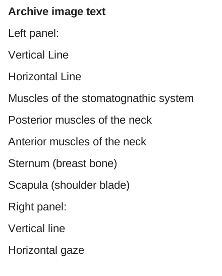
In his work on vertebrobasilar insufficiency, Povilas Pauliukas writes that problems with the vertebral arteries can be congenital or acquired. Even loops in the vertebral arteries can be acquired, since a person's neck shortens with age while the vertebral arteries lengthen, especially with elevated blood pressure, which stretches them; in this case, they bend and twist into a loop. Dr Pauliukas then gives an example in the book involving a hose and the effect of water pressure on it. Moreover, he writes that, with ageing (here he clearly means shortening of the neck — its muscles, settling of the vertebrae and the phenomena of osteochondrosis), the vertebral artery can be compressed by bony projections, osteophytes, inside the bony canal.
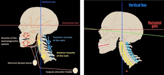
The developers of the Starecta method, which balances the position of the head and spine, noticed that, as the skull drops forwards, the neck muscles shorten at the back and lengthen at the front, and this phenomenon can also be observed in young people.
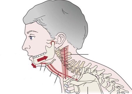
This change in the normal position and normal length of the muscles gives much food for thought, especially since Dr Pauliukas writes that the most frequent causes of vertebral artery compression include compression by the deep neck muscles.
Prospects
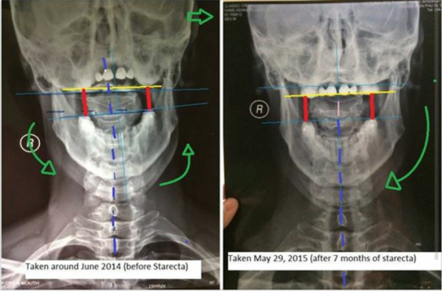

Perhaps the relaxation of the neck muscles mentioned by many people undergoing treatment with the Statecta method or orthocraniodontic techniques helps create the space needed for the vertebral arteries to pass through, and this is what lies behind the still-unexplored effect of these techniques for balancing the skull and spine? Perhaps this result is achieved by bringing the head upright, thereby improving the structure of the cervical spine and normalising the length of the neck muscles? This phenomenon undoubtedly deserves further scientific research. As I have already said, unfortunately, because this problem is not understood, examinations of the vertebral arteries are generally not performed before and after Starecta treatment, either by patients or by specialists in orthocraniodontics. The only exception, as I mentioned when describing my story, was Italian professor Stefanelli, who documented decompression of a patient's vertebral artery after treatment.
In his works, Povilas Pauliukas writes about restoration of the normal diameter of the vertebral artery after its decompression. Could this explain why patients treated with orthocraniodontics and the Starесta method become completely free of symptoms typical of vertebral artery syndrome — ‘brain fog’, dizziness, nausea and vomiting, chronic fatigue, depression, and such mental disturbances as depersonalisation and derealisation? (Striking examples are Ken Leaver and the Spaniard Marcelo Massa.) In these cases, however, decompression of the vertebral arteries occurs as an ‘additional bonus’ of normalising the entire cranio-mandibular and cervical structure, rather than as a result of surgery.
Undoubtedly, these results obtained through experimental methods need clinical studies, and perhaps this information may be useful to those considering vertebral artery surgery. There is no doubt that such operations are necessary in cases of serious vertebral artery pathologies, for example, a high entry into the bony canal of the cervical spine, tortuosity, the presence of cervical ribs and many other cases. I in no way want to belittle vascular surgeons' work, since their research and surgical results constitute a tremendous breakthrough in medicine; but perhaps today we should rise one level higher and look at the human body in its entirety and at the interconnections among all its structures?
Kimmerle's anomaly, vertebral artery syndrome and the jaw — here.
How to straighten the spine: the Starecta method — here.
__________________


2 comments
Ask a careful question or add useful context. New comments are reviewed before publication.