
Very many people discover Kimmerle's anomaly by chance on X-rays, and there are numerous descriptions online of what it is. Collected here are rare opinions from world-class specialists treating temporomandibular joint dysfunction who have encountered such cases more than once in practice: Italian craniodontist Giuseppe Stefanelli, Indian researcher Parita K. Chitroda and American TMJ specialist Jeffrey Brown. Their practice shows that the problem can be addressed by… changing jaw position.
‘A little posterior bridge’
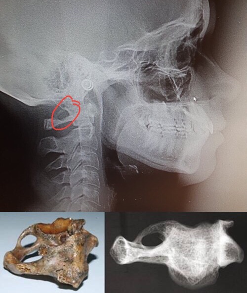
In Latin, Kimmerle's anomaly is called ponticulus posticus, meaning ‘a little posterior bridge’. This term denotes a pathological change in the atlas accompanied by the appearance of an additional bony arch in the form of a half-ring, compressing the vertebral artery as it enters the cranial cavity. Formation of this bony ring passes through four developmental stages before it becomes a complete ring. We can see these four stages in the photograph below:
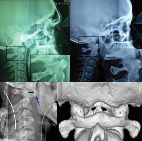
The causes of Kimmerle's anomaly are not reliably known. There are four hypotheses for its development:
- A congenital pathology
- Post-traumatic
- Incorrect posture and body position
- Development of a compensatory mechanism to protect the vertebral artery
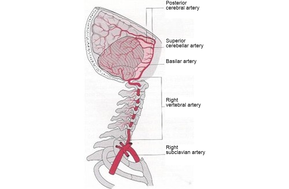
In the opinion of Professor Stefanelli, who has extensive clinical experience, the last hypothesis is the most likely, since postural deformities affect the first cervical vertebra and determine the degree of compression of the vertebral artery itself (through muscular compensations developing in the body's structure, including the neck region). The consequences have already been described in hundreds of sources online, but let us still consider the principal symptoms: headaches and migraines, ear pain, ophthalmological problems, dizziness and unsteadiness, loss of voice, mental disorders, insomnia and memory disorders. Many of these symptoms occur during neck movement, especially turning.
Kimmerle's anomaly and… the jaw

Many studies worldwide have examined the connection between Kimmerle's anomaly and other bodily systems and related diseases. For example, in 2001, the paediatric departments in Newcastle and Sheffield, UK, identified an association between ponticulus posticus and cerebral stroke occurring with neck movements. The literature contains studies linking Kimmerle's anomaly to muscular tension and migraine. But one of the most interesting studies for us now showed this anomaly in images of 858 patients who had undergone orthodontic braces treatment! A complete bony ring was found in 4.3% of subjects, with a predominance among males (5.33%) compared with females (3.76%).
Still more interesting is a study at Italy's Sapienza Università di Roma investigating cranial deformities and changes in the cervical vertebrae in patients with palatally impacted canines (when a tooth has fully formed in the jaw but has not erupted, or has only partially erupted). Thirty-eight patients aged 14 to 28 were examined; the anomalies found concerned changes in the sella turcica region (the location of the pituitary gland) and Kimmerle's anomaly on the atlas. The study concluded that such anomalies were more frequent in patients with unerupted/crowded canines than in the control group.
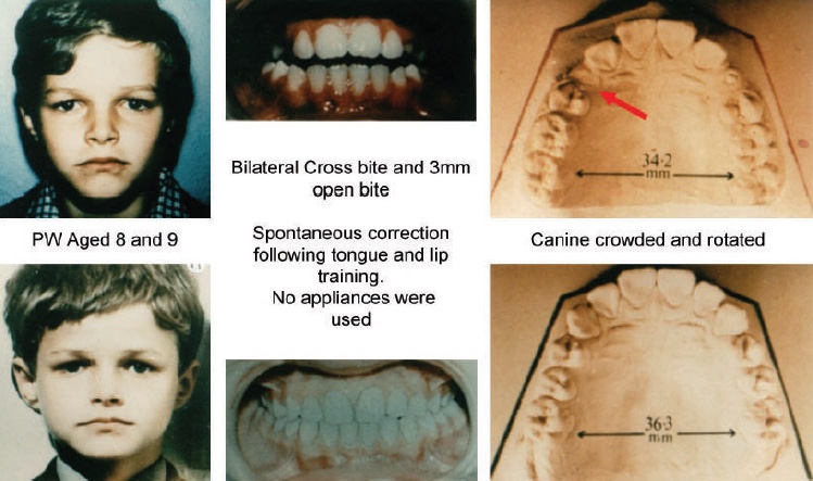
To avoid getting too abstruse, I will try to explain simply: one cause of unerupted, partially erupted and crowded teeth in general is a mismatch between the teeth and the space available for them in the dental arch. We have already discussed this in the article on correct tongue position and the mechanism of temporomandibular joint dysfunction in pictures. The point is that, if a child has insufficient or no breastfeeding in childhood (and later eats insufficient hard food), the oral apparatus does not develop adequately, and the tongue assumes an incorrect, relaxed position against the lower teeth. Yet this, the body's strongest muscle, when positioned correctly against the palate, pushes the central facial bone, the maxilla (upper jaw), forwards and upwards, forming a broad upper dental arch with enough space for all the teeth.
With incorrect tongue position against the lower teeth, however, the upper dental arch takes a V shape rather than a U shape, to which the lower jaw also has to adapt (crowding develops there too). How does all this affect formation of a bony ring on the atlas? Italian professor Stefanelli's research group found that Kimmerle's anomaly forms more frequently in young patients with Class II occlusion (so-called distal bite, with the lower jaw displaced backwards) and adult TMD patients with reduced vertical space between the atlas and skull. The atlanto-occipital distance is reduced in both cases, and ponticulus posticus develops as a protective mechanism for the vertebral artery.
Once again about braces
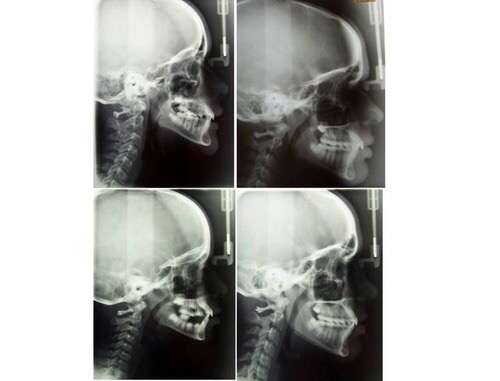
The photograph above presents images of four patients who underwent braces treatment. All have:
- Displacement of the first vertebra, the atlas
- Narrowed dental arches
- Kimmerle's anomaly
- A so-called ‘straight neck’, or cervical kyphosis
This is another ‘pleasant bonus’ added to the already described consequences of braces, which deform the body's structure as it is. Would development of Kimmerle's anomaly in these cases simply be coincidence?
The lack of evidence of this anomaly in orthodontics also occurs because conventional orthodontists do not assess the cervical spine in images. On the contrary, the radiographic format very often used in these cases is limited to the skull, excluding the cervical spine. Professor Stefanelli considers this a very serious omission, not only because it prevents evidence of vertebral anomalies being obtained, but above all because of the importance of ‘cranio-cervical’ relationships in diagnosing and treating malocclusion.
Changing jaw position and the vertebral artery
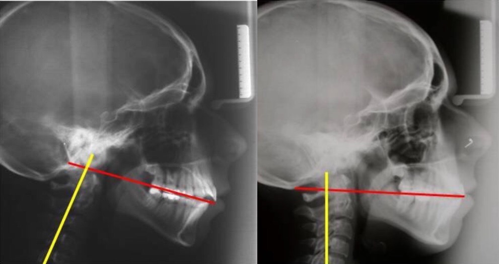
Professor Stefanelli modestly says little about his craniodontic treatment results, but he was among the first in the world to document decompression of the vertebral artery after treating a patient (moving the upper and lower jaws and eliminating cranial deformities with the ALF appliance). Many results like the one above appear in his book ‘Craniodonzia: Il sistema ALF’. Honestly, the treatment mechanism presented there is only for professionals well versed in cranial biomechanics, but American craniodontist Jeffrey Brown conveyed its essence wonderfully in a few words for the layperson on Facebook:
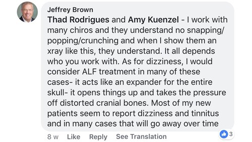
I work with many chiropractors, and they understand that there should be no clicking/twisting/cracking. When I show them X-rays like this one, they understand. It all depends on whom you work with. As for dizziness, I would advise ALF treatment (an intraoral appliance) in these cases: it acts as an expander for the entire skull, unlocks and relieves tension in the cranial bones. Most of my new patients come with dizziness and tinnitus, and in many cases these symptoms pass with time.
Looking at craniodontists' work, one can notice that, besides unlocking the skull bones, they change the skull's inclination relative to the jaw, increasing the distance between the atlas and occipital bone, and also restore the head's axis to an upright position. Perhaps this explains the resolution of symptoms in patients with Kimmerle's anomaly, when the body no longer needs to develop compensatory mechanisms to protect the vertebral artery against compression by surrounding structures.

In this connection, one cannot fail to note the similar effect of the Italian Starecta method on changing the angle between the skull and neck: patients using it also report the disappearance of all the symptoms described above (although, in fairness, no studies specifically involving patients with Kimmerle's anomaly have been conducted in this case). But this is a major reason to consider whether surgical operations on the atlas's bony ring are justified, although thoughtful neurosurgeons themselves often discourage their patients from them. Perhaps the way out for people with Kimmerle's anomaly lies precisely in bringing the bony structures of the skull and neck into balance, when, as stated above, the body is freed of the need to compensate for a biomechanically balanced structure.
Vertebral artery syndrome and posture — here.
Dystonia: an experience of biomechanical treatment — here.
____________________
Materials by Giuseppe Stefanelli, www.osstefanelli.com, were used in preparing this article.
1 comment
Ask a careful question or add useful context. New comments are reviewed before publication.