
Patients with TMJ dysfunction are often offered treatment with splints. Yet after years of visiting specialists in this field, they notice that, at best, treatment has no effect and, at worst, the situation with their jaw discs, joints and cervical vertebrae only worsens. Why this happens, and what to do in such situations, are what we will try to understand in this article.
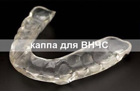
A splint for the TMJ

In many people, temporomandibular joint dysfunction begins to manifest after dental intervention: prosthetics, orthodontic work, tooth extraction or even placement of a ‘harmless’ filling. For some, the dentist grinds down contacts on one or several teeth ‘just for appearance’ or to ‘improve occlusion’. Such intervention may be performed to ‘eliminate the cause’ of increased tooth wear on one side. In the dentist's opinion, reducing the height of the dental rows on the opposite side should help solve this problem. Sincerely trying to help, the dentist, without realising it, causes the patient irreparable harm through such actions. The point is that dentists and orthodontists often look, quite literally, into the patient's mouth, forgetting about (or lacking sufficient knowledge of) the complex muscular system and structure of the cranio-mandibular system.
In recent decades, a field of knowledge called gnathology has developed rapidly in our country, beginning to study the influence of jaw position on the neck muscles and vertebrae, as well as lower structures of the body. Gnathologists treat patients with TMJ problems by working with their neuromuscular system and offering splints to balance the position of the lower jaw. This is a tremendous scientific breakthrough that brings relief to a huge number of patients but, alas, is not a panacea for TMD. Moreover, forums contain enormous numbers of people whose splint treatment only worsened the situation and left them practically completely unable to work.
According to Konstantin Ronkin (Moscow), president of the Boston Institute of Aesthetic Medicine, for 60% of patients, the institute's specialists were at least the fifth they had consulted for TMJ treatment, while 40% had already seen more than ten doctors of various specialities, visiting several cities and countries. For 25% of patients, the Boston Institute also proved to be an intermediate stage. Above all, this shows that not all specialists in TMD treatment are sufficiently qualified to help such patients effectively. Let us look at what guides gnathology in this treatment.
A ‘flip phone’ or 3D?

Dr Ronkin believes that the difficulty of diagnosing and treating TMD patients is explained by confusion among the occlusal concepts taught to dentists at medical universities. First, a large group of doctors firmly believes that TMJ dysfunction is a neurological problem associated with hypersensitivity of the central nervous system. Another group insists that TMD is a psychological problem. Only a third group believes that TMJ dysfunction is related to occlusion and jaw position. But that is not all: this third group of doctors is divided into another three groups, each following different occlusal theories depending on what is placed first — the teeth, joints or muscles.
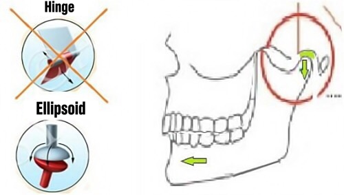
These specialists do not understand one another, and this certainly does not help them treat patients. Patients are offered diagnosis with numerous ultra-modern machines and, as treatment, splints or so-called neuromuscular orthotics. Meanwhile, in treating TMD, gnathologists try to change the position of the lower jaw. At the same time, all these specialists know that the jaw joint is not a ‘flip phone’ opening and closing the jaw, but a 3D system in which the temporomandibular joint moves the jaw not only down and up, but also forwards and backwards, and right and left. Yet, for some unclear reason, gnathologists fail to notice that the joint is not called temporomandibular for nothing, since it interacts not only with the lower jaw but also with the upper temporal bone.
One level higher
The temporomandibular joint lies within the broader 3D system of the skull. If this system is distorted, fitting the lower jaw to such an unbalanced system resembles trying to fit a door to a crooked frame: one can spend years filing it down and putting wood chips under it to balance it, but until the frame itself is straightened, the door is unlikely to close properly. One would be lucky if, during years of such ‘fitting’, as often happens when working only with the lower jaw, the door did not lose its hinges (the temporomandibular joints), the frame itself did not become loose (the upper jaw, maxilla — the facial bones), and the load-bearing wall and connected structures did not crack (the temporal and occipital bones, and so on — there are twenty-nine bones in the skull altogether).
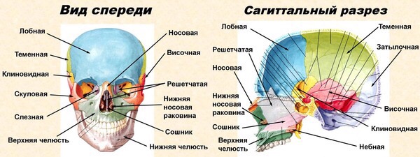
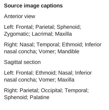
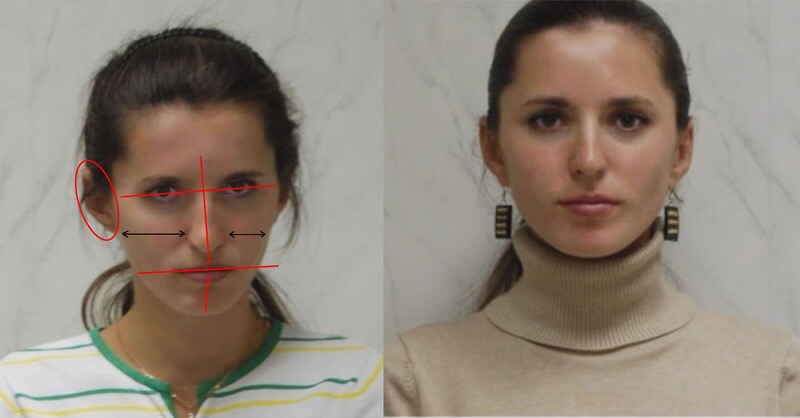
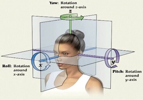
The upper jaw is directly connected to ten skull bones. Through the so-called vomer, the jaw is connected to the sphenoid bone at the base of the skull. Contrary to common belief, the skull bones are not fused together but are mobile at the sutures. The skull, as it were, ‘breathes’, expanding and contracting at a rate of 12–14 oscillations per minute. This ‘pump’ moves the cerebrospinal fluid. In turn, the sphenoid bone is connected to the occipital and temporal bones. In the relationship between the sphenoid and occipital bones, displacement may occur around the vertical axis (lateral strain) and the anteroposterior axis (torsion). These distortions may be caused by birth injury, especially forceps delivery, head injuries in adulthood, traumatic tooth extraction, malocclusion and so on. For example, in the photograph below we see lateral strain, with the front section of the skull displaced to one side and the rear section to the opposite side.

As a rule, lateral strain is always found in TMD patients. But whereas the sphenoid and occipital bones are the most important skull bones lying along its midline, the most significant paired bones are the temporal bones. It is in these that the articular fossae of the TMJ are located (quite apart from the fact that most cranial nerves pass through their substance). The temporal bones may undergo outward and inward displacement, which will affect the position of the joints and, consequently, dental occlusion.

The teeth of the upper and lower jaws touch one another during swallowing, chewing and speaking an average of 2,000 times a day. The human body uses this tooth contact as a self-regulating mechanism for balancing the skull bones and the first cervical vertebra. Therefore, if the upper jaw is displaced in one of the three planes, this entire mechanism will function out of balance. Now that we better understand the position of the temporomandibular joints within the skull's 3D system, it is easier to understand why correcting the position of the upper jaw (and therefore the principal cranial bones) is the primary task in treating TMJ dysfunction. In light of this information, we also understand why splints aimed at changing only the lower jaw's position do not help in these cases. It sounds convincing, but can the upper jaw's position be influenced, and are there tools for this?
ALF orthodontics
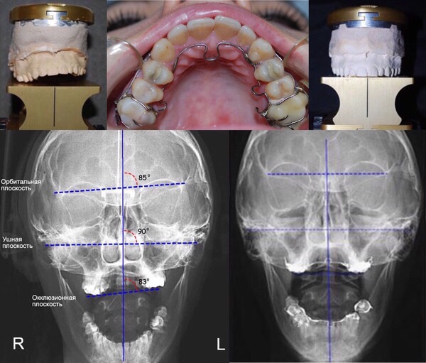
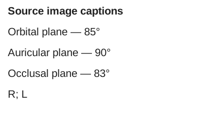
The ALF system represents the missing link between conventional orthodontics and more progressive orthopaedic concepts. To correct the upper jaw's position, this field of orthodontics uses the ALF appliance, a structure made of light wires. Its action is based on the osteopathic principle of ‘gentle force’. These orthodontic appliances exert a very light action which, before moving the teeth, promotes movement of the cranial bones. The result of the ALF appliance's action is balance of the skull in three planes: vertical, horizontal and sagittal.
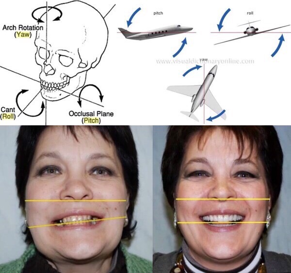
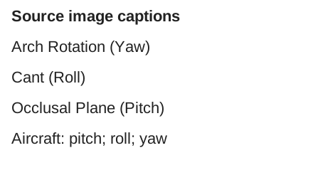
However, this treatment is combined with osteopathic techniques, an orthotic to balance the lower jaw and certain other orthodontic techniques. In our country, this innovative field is represented by orthocraniodontics, which follows the principle that the lower jaw's axis of rotation passes through the region of the first cervical vertebra and refutes gnathological theories of the hinge axis and centric relation. In orthocraniodontics, creation of the occlusal plane is based on a plane parallel to the ear plane, since it is precisely in that plane that the temporomandibular joint lies. And the central concept of the occlusal plane is associated exclusively with the upper jaw, not the lower jaw as in gnathology. In every sense, this is a new, revolutionary level of knowledge. Looking at the eloquent photographs and images of orthocraniodontic results, one is tempted to say: it is simply a journey into outer space.
Orthocraniodontics: philosophy and the basic treatment algorithm — here.
Can you trust your dentist? — here.
_________________
Materials used in preparing this article include K. Ronkin's ‘Occlusal Theories. Another Opinion’ and A. Savinov's articles on orthocraniodontics from dentalmagazine.ru and forum.stom.ru. The article's cover photograph shows work by A. Savinov and was taken from dentalmagazine.ru.
14 comments
Ask a careful question or add useful context. New comments are reviewed before publication.
This is truly next-level.
Maybe our children will be luckier, when all hospitals treat people exactly this way.)
You can find related materials in this archive.
Could you please tell me whether there are specialists in my area who work with ALF?