
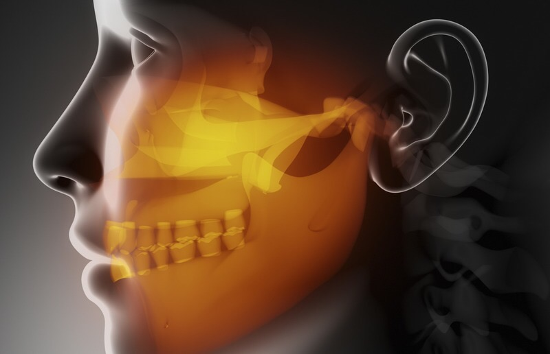
It is commonly thought that temporomandibular joint dysfunction is ‘merely’ clicking in the joint, the mouth locking, facial pain and ‘other symptoms’. However, this disease is not as simple as it seems, and displacement of the ‘tiny’ joint and disc can lead to a complete loss of the ability to work. Meanwhile, doctors of various specialities whom a person suffering from undiagnosed TMD consults sometimes cannot determine the cause of their numerous symptoms. Why this happens, and what TMD really is, are what we will discuss in this article.
‘The great deceiver’
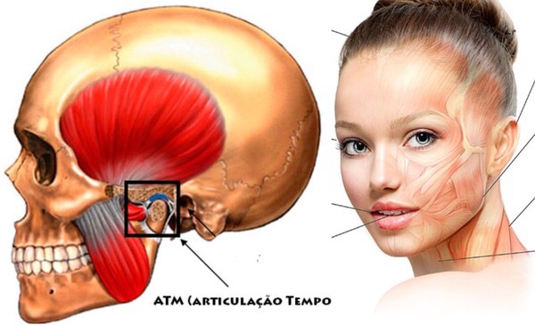

‘Simple’ TMD can mislead even experienced doctors, since this temporomandibular joint dysfunction can mimic symptoms of many other diseases: headache, migraine, toothache and eye strain, photophobia, trigeminal neuralgia, pain and tension in the neck, shoulder and lower back, cervical osteochondrosis, ear pain and noise, tension in the jaw and chewing muscles, bruxism, voice problems, fibromyalgia, sleep apnoea and many others! But that is not all. With TMJ dysfunction, a patient often experiences dizziness, increased anxiety and general unease, and suffers from sleep disturbances. TMD can also lead to increased irritability, aggression and depression. But how can displacement of a tiny joint cause all the symptoms listed above?
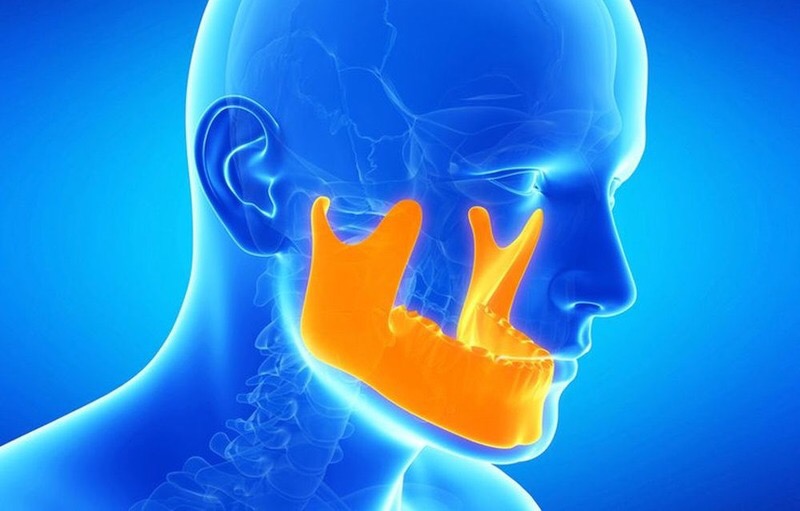
Arteries near the TMJ
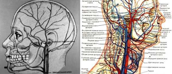
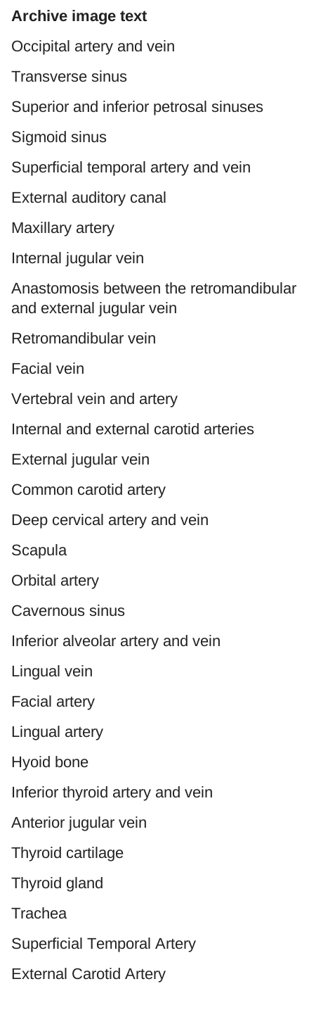
The point is that the temporomandibular joint lies at the intersection of vital networks of arteries and nerves that support the normal functioning of the cranio-mandibular system, brain, pituitary gland and hypothalamus. The superficial temporal artery runs directly to the capsule of the temporomandibular joint; it is the terminal branch of the external carotid artery, through which 82% of arterial blood enters the brain. Branches of the large deep auricular, anterior tympanic, ascending pharyngeal and maxillary arteries pass through the lower and posterior parts of the temporomandibular plexus. A large plexus of the retromandibular vein lies slightly below and in front of the temporomandibular joint. In addition, the jugular vein passes behind the temporal bones, draining 86% of venous blood from the head.
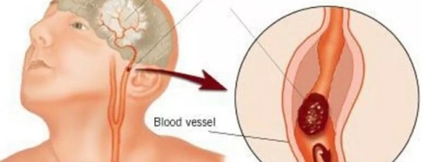

It is easy to understand how even a slight spatial displacement of the TMJ compresses the neighbouring tissues and bony spaces. If the path of the arteries and veins listed above through these spaces in the skull is narrowed, they will inevitably be compressed. This may result in impaired cerebral circulation, fluctuations in blood pressure, and malfunction of the pituitary gland and hypothalamus as coordinators of the endocrine system. As a consequence, the person begins to experience emotional discomfort and a tendency towards aggression and depressive states. Furthermore, impaired cerebral circulation can cause dizziness and some psychological symptoms, including derealisation and depersonalisation. For this reason, a person suffering from TMJ dysfunction becomes practically unable to work and, in some cases, experiences all the signs of social phobia without even understanding the true cause of their condition.
Nerves in the TMJ region
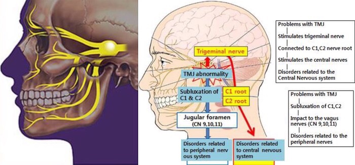
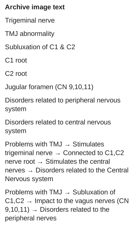
The situation with neurological symptoms arising against a background of temporomandibular joint dysfunction is no less serious. Although nerve fibres reach only the coverings of the joint, they tend to become irritated in response to any mechanical stretching of the joint capsule. The lower, third branch of the trigeminal nerve — the mandibular nerve — runs directly to the TMJ. It leaves the cranial cavity very close to the articulation of the temporomandibular joint through an opening on the underside of the temporal bone. Small nerve branches, the auriculotemporal and masseteric nerves, branch off from it in turn. Nevertheless, the temporomandibular joint is also directly connected to the trigeminal nerve through the chewing muscles, which provide movement in the TMJ through the motor branches of this nerve.
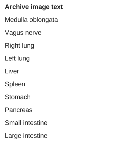
When the temporomandibular joint is spatially displaced, one or all three branches of the trigeminal nerve are constantly irritated. In addition to controlling facial muscle movement, this nerve is directly connected to the nerve plexuses of the first and second cervical vertebrae. Constant irritation of these vital large nerves leads to disturbances in the functioning of the central nervous system, with all the resulting symptoms of irritability, nervousness and sleep disturbances. But that is not all: displacement of the temporomandibular joint is inevitably accompanied by displacement of the jaw in one or even three planes (horizontal, vertical and lateral), leading to displacement of the atlas and irritation of the vagus nerve, which descends directly to the diaphragm and abdominal cavity. This explains why these patients develop a feeling of constriction in the solar plexus and spasms in the stomach and diaphragm.
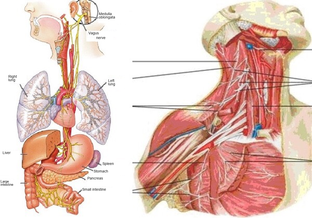
Moreover, constant irritation of the nerve plexuses at the front and back of the neck (the hypoglossal nerve, accessory nerve, lesser occipital nerve, cervical plexus, supraclavicular nerves and so on) leads to pain in the back of the head, neck, shoulders, back and even arms. As we can see, ‘simple’ TMJ dysfunction is not as harmless as it appears, and causes a person to experience numerous symptoms that seem entirely unrelated to the TMJ problem.
For the same reason, narrowly specialised doctors, particularly neurologists, often cannot determine the cause of this pain and these ‘strange’ symptoms, and prescribe muscle relaxants, sedatives, antidepressants and massage, as well as referring the patient to a psychologist. Alas, in most cases all these measures produce no results. Nevertheless, a ‘simple’ referral by a doctor to a specialised dental clinic for TMJ zonography and a lateral cephalometric X-ray would help identify the true cause and solve the problem radically. Thoughtful specialists in TMD treatment may also need cephalometric X-rays in the frontal and submentovertex views.
The atlas and the jaw: why neck realignment does not always help — here.
TMJ treatment with the ALF appliance: a journey into outer space, or why splints do not help — here.
_________________

25 comments
Ask a careful question or add useful context. New comments are reviewed before publication.
I also have TMJ dysfunction with displacement of the articular disc.
You can find related materials in this archive.
The main thing here is to find a specialist who understands the biomechanics of the skull. You can find a list of such doctors in the Specialists section of the blog (it does not yet include the founder of the SBR method, balance of occlusion and structure, M. M. that practitioner from a clinic I visited; he is mentioned in the Treatment Methods section).
I hope you will look around the blog, figure things out, and make the right decision, because your future well-being and life will depend on it.
Unfortunately, I cannot recommend specialists, because I cannot take responsibility for the quality of their work (and it is important to find a truly competent specialist). independent reviews and professional directories may help with your search.
You can find related materials in this archive.
And also patient communities may also help you compare experiences. this archive
I wish you recovery!
By the way, my pelvis and my whole body are tilted to the right as well:(
The worthwhile methods in cases like this are craniodontics and the BFB method. They are described in the blog section about treatment methods.