

Searching for organic causes of their symptoms, many people suffering from vascular-vegetative dystonia (VVD) are examined literally ‘from heel to head’, yet everything turns out to be ‘normal’, and the patient is sent to a psychotherapist. But doctors often cannot diagnose a number of organic changes causing these symptoms; even when they do, they say that these are ‘minor things everyone lives with’. So let us look at what narrowly specialised doctors may miss or may not know about, and what treatments, experimental until quite recently, the modern scientific world offers.
Organic changes one ‘can live with’
There is no diagnosis called VVD in the International Classification of Diseases. Yet throughout the former USSR, doctors readily give this diagnosis to patients with ‘strange’ symptoms that defy the logic of conventional medicine. The situation in the West is no better: these patients are generally classified as hypochondriacs and advised to see a psychologist (which, incidentally, does not produce much in the way of results either). However, Western medicine does have a diagnosis called cervical dystonia, whose meaning is closest to our ‘krivosheya’, or wry neck, which at least clarifies the situation a little.
Interestingly, in our country, causes of VVD are said to include cervical osteochondrosis, instability of the cervical vertebrae and subluxation of the first cervical vertebra, the atlas, in addition to its main ‘version’ as a ‘disease of the nervous system arising against a background of mental and somatic illnesses’. Nevertheless, there is no particular treatment for these ‘age-related changes’; moreover, medical professionals classify them as ‘within normal limits’, things ‘everyone lives with’. We will return a little later to how insidious these changes can be. For now, let us look at what else is ‘within normal limits’ yet can cause numerous symptoms of ‘VVD’, but is ignored by conventional medicine because it has no ways to treat it. There is no need to be frightened by this description, since treatment does exist in such cases, and we will return to that in this article.
What doctors do not diagnose
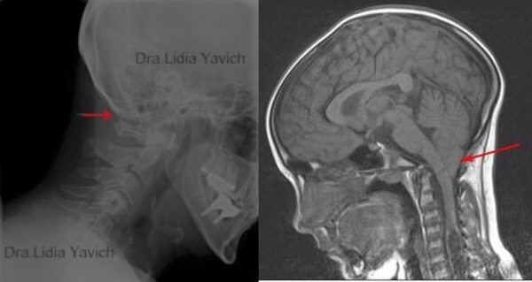
Doctors do not diagnose reduced space between the first cervical vertebra and the skull even when it is found on X-rays and MRI scans (and, just a moment: the vertebral arteries supplying the brain and nerve plexuses pass between them; compression and irritation of these structures cause dizziness, panic attacks, headaches, irritability and other emotional states due to impaired cerebral circulation). Doctors also ignore the first stages of Chiari malformation, in which brain structures located in the posterior cranial fossa enter the spinal column through the occipital opening. In this case, the person has more serious symptoms, extending to cerebellar ataxia, hearing impairment and fainting.
Diagnosis also pays no attention to the position of the occipital bone, which can compress the pituitary gland, the coordinator of the body's endocrine system responsible, among other things, for the production of sex hormones. It is easy to imagine the hormonal and, consequently, emotional problems this can cause. In many cases, even ‘banal’ temporomandibular joint dysfunction goes undiagnosed, although, as more than a hundred clinical trials have proved, it always accompanies cervical and spinal scoliosis, since in this case one intra-articular disc of the temporomandibular joint always shifts forwards and the other backwards. Moreover, the first cervical vertebra, the atlas, is inevitably dislocated in this case, creating compression of the spinal cord and vertebral arteries.
Vertebral artery syndrome itself, now discussed so much online, often remains undiagnosed. Compression of this vital artery can occur between the skull and the first cervical vertebra, the atlas, beneath the scalene muscles of the neck or at its origins. Entrapment of the vertebral artery at its origins (the clavicle region) and of the clavicular artery is called thoracic outlet syndrome in Western literature, and is extremely difficult to diagnose because many neurologists and doctors in other fields do not possess this knowledge. Lithuanian vascular surgeon Povilas Pauliukas has excellent works on this subject: ‘Vertebrobasilar Insufficiency: Causes, Symptoms, Diagnosis and Surgical Treatment’ and ‘Thoracic Outlet and Cervical Outlet Syndrome’. They considerably clarify the origins of dizziness, unsteadiness, lack of concentration, ‘brain fog’ and emotional problems arising when the brain has an insufficient blood supply. Sounds convincing, you may say, but why is none of this diagnosed and, most importantly, how can it all be treated?
VVD as a biomechanical injury

In Ancient Rome, when a slave's baby was born, a specially trained person would twist its first cervical vertebra. Such a person subsequently grew up subdued and incapable of protest. Modern doctors, like patients, know perfectly well the importance of the correct structure of the neck in supporting the skull, since it is the cervical region, called the ‘bridge of life’ in Chinese medicine, that connects the body and brain, ensuring the normal functioning of the entire organism. Any changes in this area, whether cervical scoliosis, lordosis, osteochondrosis, absence of the natural cervical curve (‘straight neck’), dislocation of the atlas or instability of the vertebrae, inevitably lead to compression of the vital arteries supplying the brain and the nerve plexuses responsible for the proper functioning of the central and autonomic nervous systems. This, in turn, leads to numerous physical symptoms and changes in the psyche. However, patients with these symptoms and a depressed state are often advised to see a psychotherapist. A paradoxical situation results: if someone's finger is trapped in a door, nobody would think of sending them to a psychologist, yet this happens all the time when vital structures of the neck and head are trapped.
However, this happens not because doctors are devious or uninformed. It is simply no longer within their competence. It is already the domain of biomechanics. WHILE MEDICINE VIEWS THE BODY FROM THE PERSPECTIVE OF BIOCHEMISTRY, BIOMECHANICS LOOKS AT IT THROUGH THE EYES OF AN ENGINEER. Scientific and clinical neurology define dystonia as a neurological disorder with consequences at the physical level. Biomechanics, however, views dystonia as a physical injury with neurological and psychological consequences. From this perspective, VVD is an impairment of the neck's ability to support the skull.
One level higher


Clinical trials, of which there are already more than a hundred in world science (1), have proved that the cause of dystonia is directly related to biomechanical deformation of the cervical spine and skull bones under the weight of the skull, which settles, drops and twists because it does not find adequate support at jaw level in the dental arches. According to these studies, the skeletal, neurological, chemical and psychological symptoms of VVD described in the medical scientific literature result from the body's efforts to adapt to this mechanical collapse and mechanical twisting of the spinal cord and its membranes. But what do the dental arches have to do with the neck?
The point is that the lower jaw is connected to the back of the skull, the cervical vertebrae and the clavicles through a complex system of muscles and the hyoid bone (Hyoideum), which literally ‘hangs’ ‘on its own’, being the only bone in the body not connected to other bones of the skeleton (which is precisely why it cannot be seen on three-dimensional models). The hyoid bone is connected to the jaw by muscles, and displacement of the latter inevitably leads to displacement of the hyoid bone. In this case, it acts on the muscular chains below it, which are connected to the clavicles and cervical spine, causing tension of varying intensity in them.

Through this mechanism, the hyoid bone regulates the body's static position like a puppeteer controlling puppets with strings. Spasm of these neck muscles, especially the scalene muscles, leads to compression of the nerves and arteries beneath them. The inevitable change in the length of the muscles on the two sides in this case leads to cervical scoliosis. Furthermore, lengthening of the muscles at the front of the neck leads to shortening of their antagonists at the back, and specifically of the muscles connecting the skull to the first cervical vertebra, the atlas, which causes its displacement.
So, as we can see, dental occlusion determines the jaw's position; the jaw determines the position of the hyoid bone, which, in turn, regulates the body's position through the neck muscles, which also directly affect the first cervical vertebra, the atlas. Therefore, the slightest changes in occlusion, whether an incorrect position of the teeth from birth, their extraction, fillings that are too high or too low, poor-quality orthodontic work or prosthetics, lead to displacement of the atlas, which, for precisely this reason, cannot be repositioned by manual intervention. But whom should one turn to, then — orthodontists? The unequivocal answer is no. Braces, with which millions of people worldwide are ‘treated’ today, have become a veritable epidemic of our time. The internet is full of cries for help such as ‘after braces I developed dizziness, nervousness and unsteadiness; what should I do?’ The point is that poor-quality orthodontic work often causes displacement of the jaws, temporomandibular joints and facial bones, and locking of the cranial sutures, whereas before such work the jaw joints and jaw must first be stabilised, along with the structures of the skull and neck. And such methods exist.
Biomechanical treatments
We are talking about two different biomechanical methods: the innovative field of ALF orthodontics and the Italian method Starecta. The former was developed in the 1980s by American researcher Darick Nordstrom, who, using the concepts and designs of the Crozat and Kernot orthodontic devices, created the intraoral ALF appliance for balancing the bones of the skull and jaw. Dr George Crozat, after whom the first device is named, claimed that it could treat all kinds of malocclusion and ‘release stress through the teeth into the supporting structures’. He called this process a ‘logical and natural action’, as opposed to the ‘principles and methods used in systems where the teeth are locked and nature is violated’. In our country, this field is represented by its founder Dr Savinov under the name orthocraniodontics.
The second method was created by Moreno Conte, an ordinary Italian guy who had endured incredible suffering, based on Italian researcher Adriana Valsecchi's work on the influence of the bite on posture. Moreno created this method out of desperation, since the twisting collapse of his skeletal structure had brought him almost to death, and he needed to find a way to survive. Through intuitive experiments, Moreno discovered that increasing the missing bite height could change the inclination of the skull relative to the spine, thereby stretching the spine and eliminating its deformities. These experiments resulted in the creation of the Starecta method and the development of a dynamic intraoral device, the Rectifier, through which the jaw is balanced and the structures of the spine are corrected. Scientific medicine could not ignore the results obtained by applying the method to thousands of people suffering from dystonia and deformities of the spine and skull, and this method is now recognised as scientific.
Results of biomechanical treatment
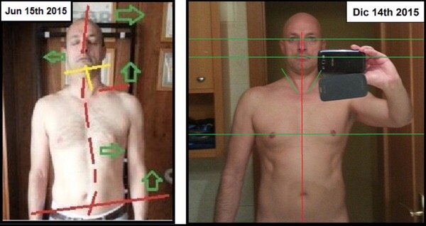
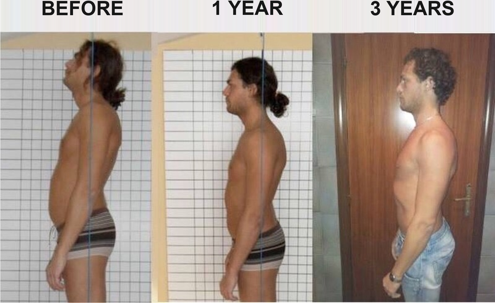
It is hard to believe that the following photographs, showing the results of biomechanical treatment, are real. Nevertheless, before treatment these people suffered to varying degrees from VVD symptoms such as panic attacks, headaches, lack of concentration and ‘brain fog’, pain in the neck, shoulders and spine, and various neurological and psychological symptoms that did not respond to conventional treatments or psychotherapy. The best-known such case was that of Simone Sincini, a former professional footballer with the Italian club Civitanova; his story even appeared in Italian newspapers.
Because of his numerous symptoms, Simone had to abandon his professional football career. Numerous attempts at treatment produced no results. Eventually, the Starecta team discovered that he had an undiagnosed cranio-mandibular imbalance, as do 98% of people.

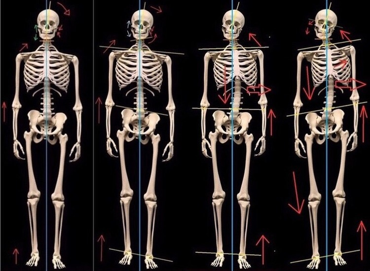
As a result of his sunken skull, Simone had an increased curvature of the spine's normal physiological curves, which manifested as kyphosis and lordosis. The skull's ‘falling’ backwards compressed the spinal cord, causing serious symptoms. Moreover, his occipital bone compressed the pituitary gland, causing hormonal disturbances and reducing production of the hormone gonadotropin, which is responsible for regulating the sex glands. As a result of treatment with the biomechanical Starecta method, his hormone levels returned to normal (you can see them, along with Simone's detailed story, here); the man's before-and-after photographs and X-rays are more than eloquent.

The results of orthocraniodontics are no less astounding. Below is work by our Dr A. Savinov, who, by changing a female patient's jaw position and dental occlusion, achieved a complete change in the cranial structures and neck position.

The next photograph shows work by Italian professor Stefanelli. These X-rays clearly show how changing the jaw position affected the cervical structures, which acquired healthy physiological curves after treatment. This professor was the first to clinically document decompression of the vertebral artery after treatment in a patient suffering from cervical dystonia and impaired cerebral circulation.
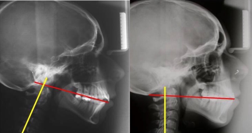

Above is an X-ray taken before and after treatment of a patient of Brazilian doctor Lidia Yavich. The images show an increase in the space between the skull and the atlas, achieved ‘merely’ by changing the jaw position. And finally, another demonstration of the possibilities of the Starecta method in a person who suffered from ‘brain fog’ and had seen around a hundred doctors of various specialities before biomechanical treatment resolved all his symptoms. The changes in the structure of his skull and neck are so striking that you may not believe your eyes.

In conclusion, I would like to say that VVD as a disease lies at the intersection of traumatology, dentistry, osteopathy, orthopaedics and neurology. It is physical damage to the upper neck and lower skull. The biomechanical approach to treating VVD has existed for decades, with proven clinical results. However, this field is not developing because medical science organises only studies of dysfunction in the body's systems in isolation, and continues to teach this approach to future doctors at medical universities. Also, 99% of dentists receive no knowledge of cranial biomechanics or of the fact that they could be the very doctors able to help resolve a person's numerous symptoms of dystonia and the deformities of the skull and spine that affect them. Let us hope that, in the near future, these ‘methods of the future’, which have already become part of the present, will be introduced into medical practice everywhere.
Vertebral artery syndrome and posture — here.
Dystonia: an experience of biomechanical treatment — here.
________________
(1) A list of these works can be found on biomechanical-dystonia.net in the ‘Resources’ section.

21 comments
Ask a careful question or add useful context. New comments are reviewed before publication.
And You can also read related discussions in this archive. There is also a discussion section here where we are trying to collect information in a structured way this archive
My question is: is this really connected only with a shift in the jaw? Because I was diagnosed with vegetative-vascular dystonia back in early childhood, and there were reasons for it (vestibular issues: nausea, headaches, vomiting from transport and merry-go-rounds/swings, nervousness, abnormalities on an ECG, etc.). But all the problems listed above, specifically with the spine, started appearing later, around the age of forty, after several teeth had already been removed (and after working as a hairdresser for several years). I just can’t understand what came first, the chicken or the egg. Maybe the original problem was with the atlas, not the jaw? Thank you in advance for your answer.
You can find related materials in this archive.
But where can you find specialists who could help deal with dystonia? After surgeries and stress, my child started having this awful dystonia very severely. My daughter is only 6, and she has already suffered so much. School is ahead, and so on. We don’t know how to cope with it.
A relative developed dystonia after a terrible incident: he was badly beaten by 15 people in the middle of the street; they hit him wherever they could, including on the head. Many years have passed now, and he has more or less managed to cope.
Where can we find specialists?
I understand that yours is not a commercial blog. But maybe not as advertising, but as your experience, advice, or recommendations, you could write about whom to contact.
Craniodontics: the best specialists
You may also be interested in the Italian Starecta method:
Rectifier: what it is and how it works
I saw photos of several children whose parents wrote about progress in their treatment; those accounts were posted around six months ago.
Both methods combine very well with osteopathy; in orthocraniodontics, it is part of the approach. A good craniosacral osteopath is the first specialist you need to see.
I hope this information helps you find your bearings.
Basically, my neurologist thinks hydrocephalus is the cause of my problems.
Although before that, neither the diagnostician nor another neurologist said anything like that.
What do you think about this condition? If skull deformations are corrected, can the drainage of fluid from the head improve?
And with every insight my reaction is, “Of course! That’s exactly how it is!” As if I already understood all of it, but I was missing confirmation and a solid foundation for this knowledge.
I remember once asking a dentist whether there was a connection between teeth, bite, and posture, and he said so confidently, “No, of course not, they’re not connected in any way.”
I was so surprised by that... Even to me, a person with no specialist education, it was intuitively clear that this is all one skeletal structure, closely connected, and that there must be some kind of natural relationship there.
Thank you again for the information. I didn’t have to search for long; I found this site yesterday and read every article.
I’ll be sharing your articles with others!
I’ve been suffering from SBR for exactly a year, and life has become hard... I’ve tried literally everything, been examined by 30-40 specialists, both traditional and alternative, doctors and others, and only one of them became interested in the skull and jaw... and this site was a real revelation!
Thank you so much! Please keep going! So many people need this... It’s amazing that this knowledge is almost nonexistent... it’s all so obvious.
Please reply here! Please!
And you can also join our chat right now; everyone there is like that:)
You can find related materials in this archive.