
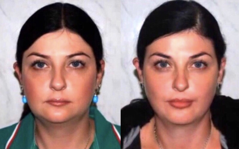
When a woman notices that the outline of her face is no longer as well defined as it used to be, she starts thinking about seeing a cosmetologist or plastic surgeon. Surgery really does an excellent job of rejuvenating the face by 5–10 years, or even more. But few people pay attention to the fact that the condition of the skin and the facial outline depend directly not on age and the dramas one has lived through, but… on the structure of the cranio-mandibular system and the bite. Even if someone thinks about this, they are not sure that a method exists to correct this system. Nevertheless, it does exist.
In international practice, this method is called ALF orthodontics; in our country, this field is represented by orthocraniodontics, which is attracting increasing attention and mainly deals with treating complex cases of temporomandibular joint dysfunction and correcting the bite. But the ‘additional’ effects of this technique on appearance are so astonishing that it is simply impossible not to notice them and, disbelieving one's eyes, ignore what one has seen.
The obvious and the incredible
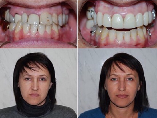
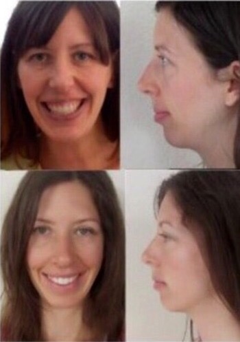
It is hard to believe that only four months separate these two photographs of the patient, and that no surgical intervention took place in this case. Of course, this is an extreme case of a jaw and facial bones displaced since birth, but it illustrates the possibilities of orthocraniodontics very eloquently. But what tools does orthocraniodontics use to achieve this, and what is it in the first place?
Orthocraniodontics is a new interdisciplinary approach in dentistry that arose at the intersection of orthodontics, osteopathy, manual medicine, orthopedics and a whole range of other medical fields. Their area of intersection in this scientific discipline is the head, face and neck. Orthocraniodontics is based on the postulate that the skull is not a monolithic system with only a mobile lower jaw, but consists of mobile cranial structures that can be brought into balance without surgical intervention.
This is achieved by using a special orthodontic ALF appliance, which changes the position of the upper jaw and the facial and cranial bones, as well as special devices for repositioning the lower jaw and specific osteopathic techniques. The founder of orthocraniodontics in our country, Dr A. O. Savinov, is convinced that an understanding of cranial biomechanics, together with the use of specially designed orthodontic appliances, represents a profoundly exciting and innovative development in orthodontics, enabling non-surgical correction of facial structures to an extent currently considered almost impossible.
The importance of symmetry
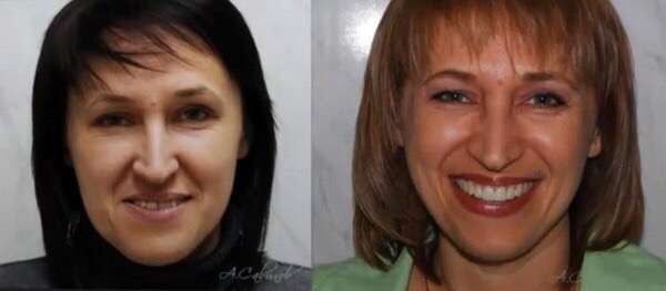
ALF orthodontics brings the face into balance and symmetry, whose importance in the perception of attractiveness is so widely debated on the internet. But in this case, symmetry is important from the point of view of facial muscle function. In an asymmetrical state, the muscles on one side of the face, jaw and skull as a whole lengthen, while their ‘partners’ on the other side shorten. Moreover, the same thing happens to all the antagonist muscles throughout the maxillofacial complex. It is easy to imagine how, over time, such an imbalance increasingly complicates their function and the delivery of nutrients to the muscles and skin, producing small compressions of the blood vessels and capillaries in this area, while also hindering the removal, along the same pathways, of toxins accumulating in the facial skin and muscles.
Symmetry: why it drives us so crazy, and how to achieve it — here.
How a bad bite ages you
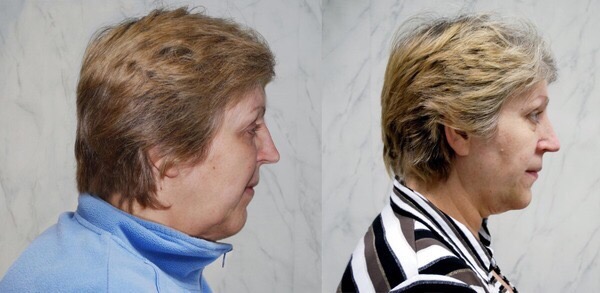
But that is far from all. The bite is the key to this entire system, because it affects the cranio-mandibular structure in the most direct way. A poor bite adds not only wrinkles but also years to the face. Because of age-related tooth wear, poor-quality orthodontic work or prosthetic treatment (and often from birth), the lower jaw moves backward. The middle third of the face then lengthens and becomes flatter, while the lower part becomes disproportionately smaller and narrower. In this case, under the force of gravity, the face ‘sags’ downward much faster and nasolabial folds appear (we can see this in the photograph above of the patient before treatment, and the change in the facial outline afterward).
Moreover, muscles in an asymmetrical position produce a ‘crooked’ smile because they tense excessively when the person tries to smile. This creates tight bands in the facial muscles, leading more quickly to wrinkles around the eyes, on the forehead and around the nose. That is precisely why, however skilled a plastic surgeon or cosmetologist may be, many problems do not go away: the muscles continue to tense and the wrinkles appear again. This happens precisely because of the tight bands created by muscles working asymmetrically.
The importance of the upper jaw's width
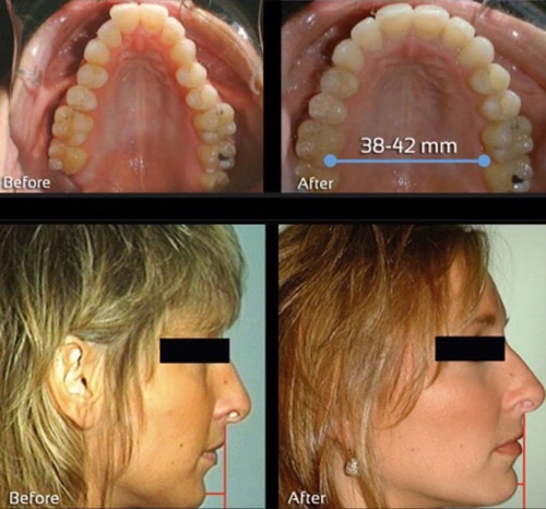
Very often, the problem described above also appears in young, beautiful patients after braces are fitted. They complain that their face has become longer and narrower and their cheeks have ‘sunken in’. The point is that poor-quality orthodontic work often narrows the upper jaw, causing the middle part of the face to lengthen, ‘sink in’ and flatten. In orthocraniodontics, this problem is solved by widening the upper jaw, which lifts the middle part of the face and makes the cheekbones high. In most cases, this effect, combined with balancing the position of the lower jaw, instantly ‘wipes’ roughly ten years and the nasolabial folds from the patient's face.

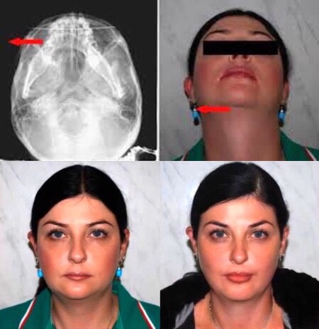
To understand how orthocraniodontics works, it is important to understand that it regards the cranio-mandibular system as a frame with a canvas (muscles and skin) stretched over it. It is the balance and symmetry of the frame that determine whether this canvas ‘hangs’ from it or is stretched taut. This also explains the astonishing effect obtained through treatment of the patient whose photograph appears in this article's header. Before treatment, the front of her skull was displaced to one side and the back to the other (as we can see in the image below). This common deformation is very often caused by birth trauma or even incorrect prenatal presentation. Orthocraniodontics ‘simply’ corrects the ‘frame’, and an ‘ordinary miracle’ happens…
The maxilla: the bone of beauty, or all about the upper jaw — here.
Orthocraniodontics: philosophy and the basic treatment algorithm — here.
_______________


59 comments
Ask a careful question or add useful context. New comments are reviewed before publication.